
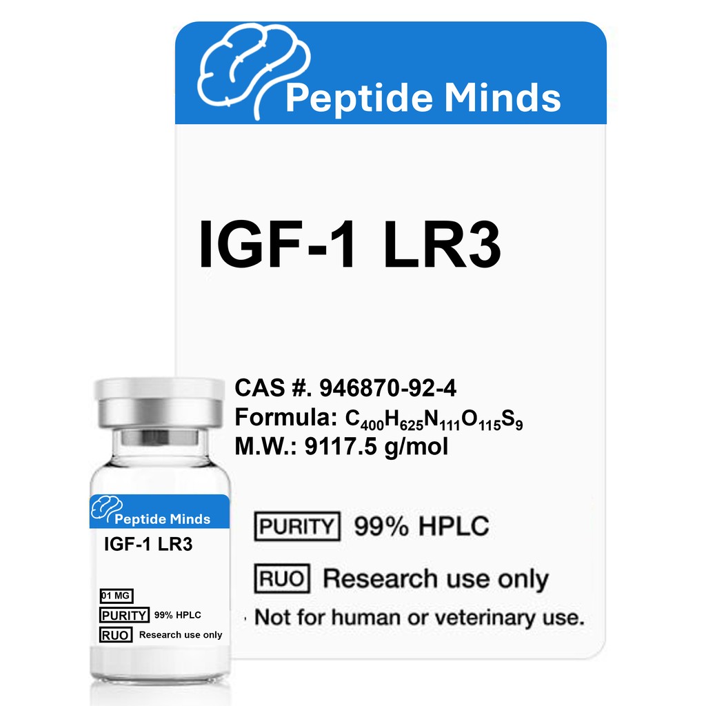
What is IGF-1 LR3?
IGF-1 LR3 (Insulin-Like Growth Factor-1 Long Arginine 3) is a synthetic peptide derived from insulin-like growth factor-1 (IGF-1), a hormone critical for growth and development. Unlike native IGF-1, IGF-1 LR3 is modified to resist binding to IGF-1 binding proteins, extending its half-life up to 120 times longer—approximately 20-30 hours versus 10-20 minutes for IGF-1. This modification involves substituting arginine at position 3 and adding a 13-amino-acid extension at the N-terminus, enhancing its potency for research. IGF-1 LR3 is widely studied for its roles in cell division, muscle growth, and fat metabolism. Learn more about peptide research.
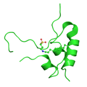
IGF-1 LR3 Structure
IGF-1 LR3 is a synthetic peptide engineered from IGF-1 with specific modifications to boost its stability and bioactivity. Below are the key structural details:
- Amino Acid Sequence:
MFPAMPLSSLFVNGPRTLCGAELVDALQFVCGDRGFYFNKPTGYGSSSRRAPQTGIVDECCFRSCDLRRLEMYCAPLKPAKSA - Key Modifications:
- Substitution: Arginine (R) replaces glutamic acid (E) at position 3 of the native IGF-1 sequence.
- N-Terminal Extension: A 13-amino-acid chain (MFPAMPLSSLFVN) is added to the N-terminus.
- These changes reduce binding to IGF-1 binding proteins, extending the half-life from 10-20 minutes (native IGF-1) to 20-30 hours.
- Molecular Weight: 9117.5 g/mol
- CAS Number: 946870-92-4
The structural modifications make IGF-1 LR3 more resistant to degradation, ideal for studying prolonged growth factor effects in cellular research. The arginine substitution and N-terminal extension are critical for its enhanced stability and activity.

IGF-1 LR3 Research
Cell Division
IGF-1 LR3 is a potent stimulator of cell division and proliferation, impacting tissues like muscle, bone, liver, kidney, nerve, skin, lung, and blood. As a key mediator of growth hormone effects, IGF-1 LR3 supports cellular growth and differentiation in research models. Its role in tissue regeneration makes it a focal point for studies on wound healing and organ repair.
Unique IGF-1
Unlike native IGF-1, IGF-1 LR3 is more potent due to its extended half-life (20-30 hours) and reduced binding to IGF-1 binding proteins. This allows for sustained activity, making it ideal for research on cellular growth, muscle hypertrophy, and metabolic regulation, particularly in models of aging and tissue repair.
Fat Metabolism and Diabetes
Studies on IGF-1 LR3 show its potential to enhance fat metabolism by promoting lipolysis and reducing adipose tissue. Research also indicates its ability to lower blood sugar levels and improve insulin sensitivity, offering insights into diabetes management and metabolic disorders in preclinical models.
Impairs Myostatin
IGF-1 LR3 inhibits myostatin, a protein that limits muscle growth. By blocking myostatin, IGF-1 LR3 promotes muscle hypertrophy and repair, making it valuable for research on muscle-wasting conditions like muscular dystrophy. Studies in animal models suggest it can reduce muscle loss and enhance recovery.
IGF-1 LR3 Longevity Research
Research in animal models, such as mice and cows, indicates that IGF-1 LR3 may support tissue repair and longevity by protecting against cellular damage and aging effects. Its prolonged activity makes it a candidate for studying anti-aging mechanisms and age-related diseases like kidney dysfunction. Discover longevity studies.
Referenced Citations
-
Adipose Tissue-Derived Stem Cell Secreted IGF-1 Protects Myoblasts from the Negative Effect of Myostatin. Hindawi. Available: https://www.hindawi.com/journals/bmri/2014/129048/ [Accessed 16-May-2019].
-
Yang, N.L.Q., Walker, R.G., Waker, T.B., Thompson, M.D., and D.B. Rodgers. Myostatin Attenuation In Vivo Reduces Adiposity, But Activates Adipogenesis. Endocrinology, vol. 157, no. 1, pp. 282–291, 2016. Available: https://academic.oup.com/endo/article/157/1/282/2422475.
-
Corpas, E., Harman, S.M., and M.R. Blackman. Human Growth Hormone and Human Aging. Endocr. Rev., vol. 14, no. 1, pp. 20–39, 1993. Available: https://academic.oup.com/edrv/article/14/1/20/2548315.
-
Sonnatag, W.E., Csiszar, A., deCabo, R., Ferrucci, L., and Z. Ungvari. Diverse Roles of Growth Hormone and Insulin-Like Growth Factor-1 in Mammalian Aging: Progress and Controversies. J Gerontol A Biol Sci Med Sci, vol. 67, no. 6, pp. 587–598, 2012. Available: https://academic.oup.com/biomedgerontology/article/67/6/587/556496.
-
IGF1/IGFBP system: Metabolism Outline and Physical Exercise – Part I. NCBI. Available: https://www.ncbi.nlm.nih.gov/pmc/articles/PMC7012221/ [Accessed 16-May-2019].
-
Hankenson, B.Y., Hancock, C.A., Peterson, C., Hornikoff, L., and J.J. Crawford. Implications of Glucocorticoid Therapy in Idiopathic Inflammatory Myopathies. Nat. Rev. Rheumatol., vol. 8, pp. 485–497, 2012. Available: https://www.nature.com/articles/nrrheum.2012.85.
-
Philippou, A., Halapas, M., Maridaki, M., Koutsilieris, M. Muscle Regeneration: Cellular and Molecular Events. In Vivo, vol. 21, no. 5, pp. 779–796, 2007. Available: http://iv.iiarjournals.org/content/21/5/779.full.
-
Philippou, A., Papageorgiou, G., Bogdanis, A., Halapas, M. Journals. 2009. [Note: This citation lacks sufficient detail for precise linking. Likely related to IGF-1 research by Philippou et al., but further clarification is needed.]
Reviews
There are no reviews yet.